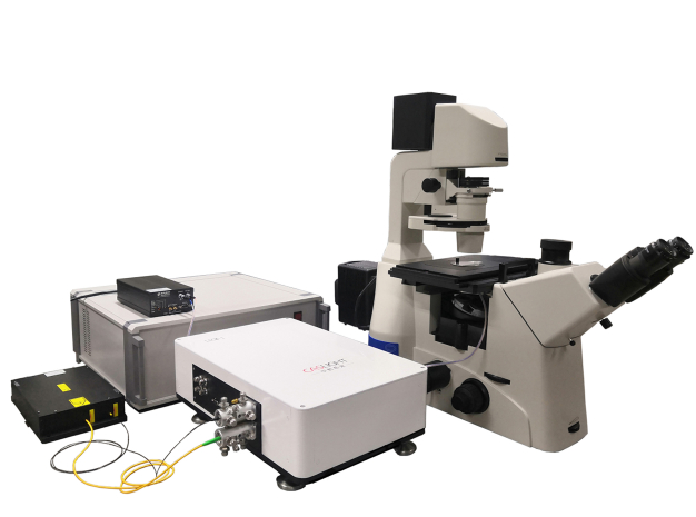
Modular design
Compatible with a variety of microscopes
Customized service
405nm、488nm、561nm,638nm,power≥20mW,can continuously adjust the laser intensity
Each laser switch and power adjustment can be controlled separately, the software can directly adjust all laser switches and the intensity
2 independent silver-plated XY scanning galvanometers
50Hz、100Hz、200Hz、400Hz、100lines/s to 600lines/s optional
128×128、512×512、1024×1024、2048×2048
220nm
XY、XYT、XYZ、XYZTetc.
Z-axis focusing
Scanning table equipped with piezoelectric objective lens focusing (travel≥300μm)
Filter optical splitting
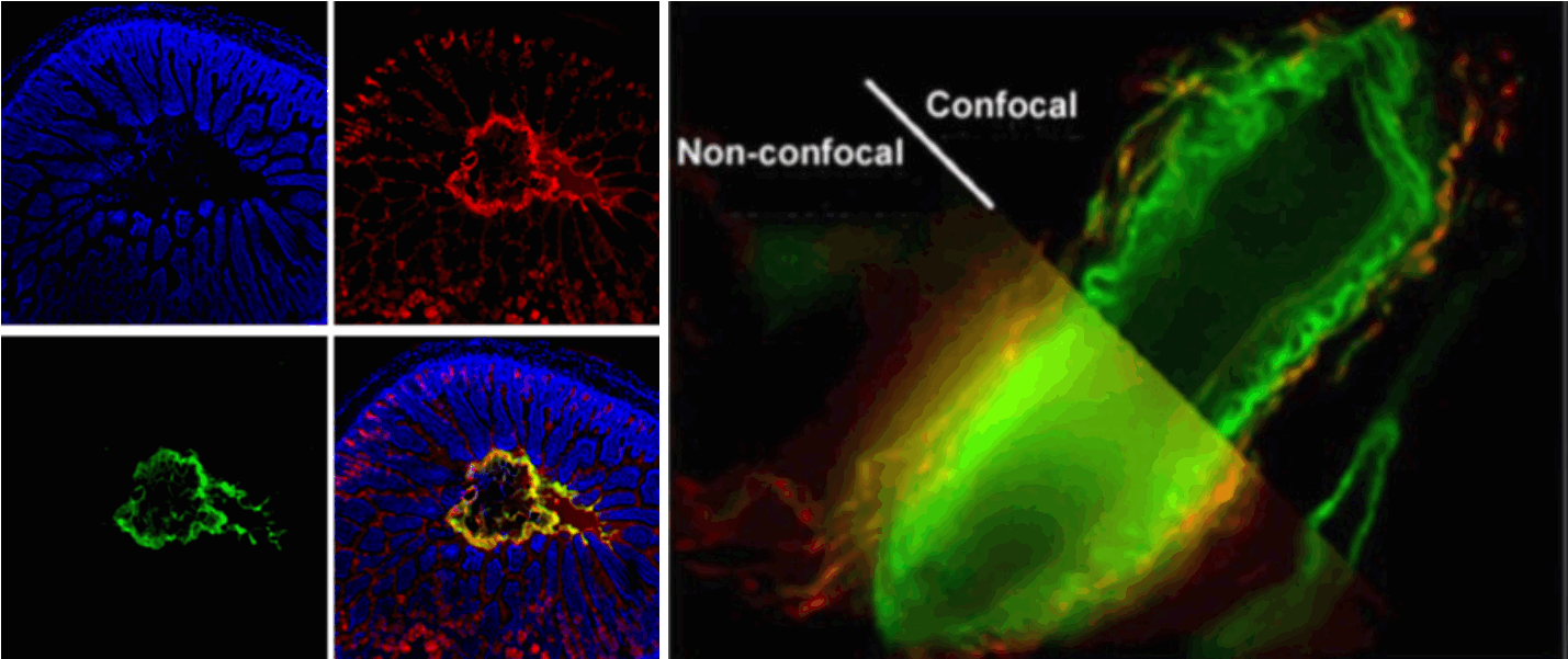
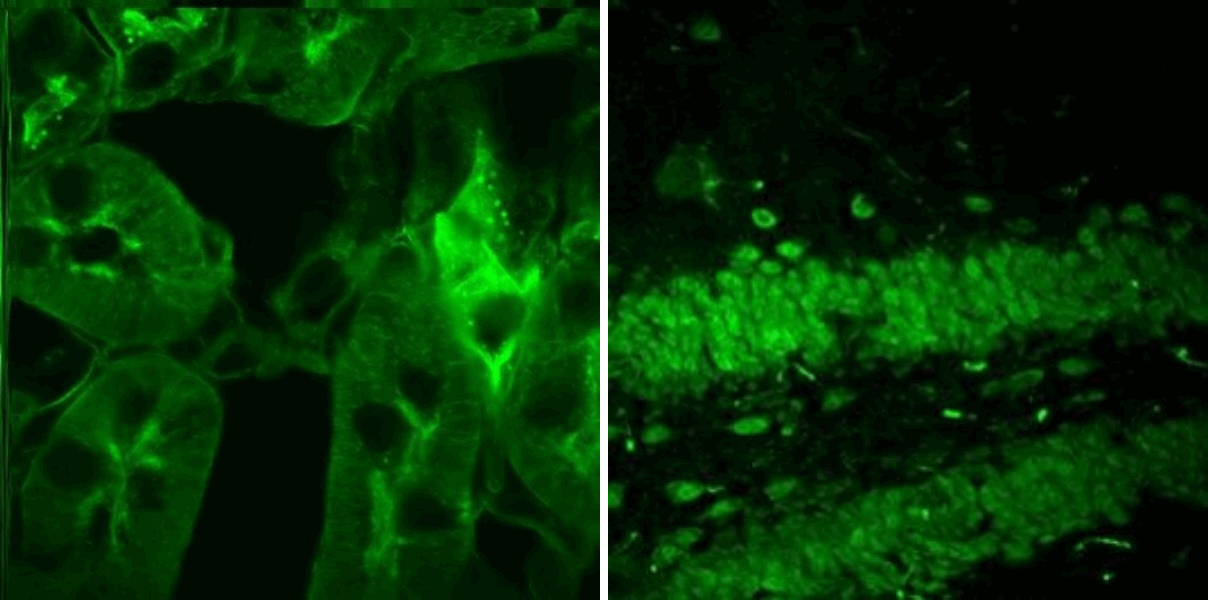
≤4 channel fluorescence detection, all channels can be scanned in real time and simultaneously superimposed
High sensitivity photomultiplier tube ( GaAsP) or Multi-alkali photomultiplier tube, the spectrum detection range is 400nm-700nm
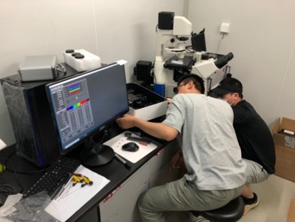
Windows10 64bit operating system
Inverted fluorescence microscope (equipped with optical plate)
10X(NA 0.42)、20X(NA 0.75)、40X(NA 0.95)、100X(oil lens,NA 1.4)APO objective lens,pair of eyepieces (10X)
30μm(about 1AU)
Graphic user interface, user setting interface, image processing interface, image acquisition and system automatic control function specially designed for dark environment
2D image processing functions, including image brightness, contrast adjustment, zooming, cropping, measurement, scale, pseudo-color disassembly, etc. It has user setting interface, image processing interface, image acquisition, automatic control system , and a graphical user interface specially designed for the darkroom environment.
3D image synthesis, observation, zoom, contrast, etc.
Preview: single-channel or multi-channel detection can be realized, and the imaging display can be performed at different speeds;
Multi-channel imaging: single-channel, multi-channel imaging and multi-channel fusion can be realized;
Multi-channel time-sharing imaging: multi-channel time-sharing imaging and multi-channel fusion can be realized;
Image saving: The scanned images can be saved in real time;
Crop function of scan field: could select the imaging area of interest for imaging;
Rotate function of scan field (manual);
Time series: Automatic imaging of the sample at a customized time interval;
Z Stack and time series: Automatic Z-axis tomography of the samples with customized time intervals;
Image processing.
Kunming Institute of Zoology - Chinese Academy of Sciences
Institute of Neurology - Suzhou University Shanghai Institute of Materia Medica - Chinese Academy of Sciences Institute of Zoology - Chinese Academy of Sciences
Modular design:
Easy to install and maintain, the purchase of modules can directly upgrade the experimental microscope
Suitable for a variety of microscopes:
Nikon, Olympus, Yongxin, etc
Customized services:
Customization can be carried out according to customers' needs so as to meet special needs such as scientific research and clinical practice.

 Download:
 Scientific Research Instruments